Introduction
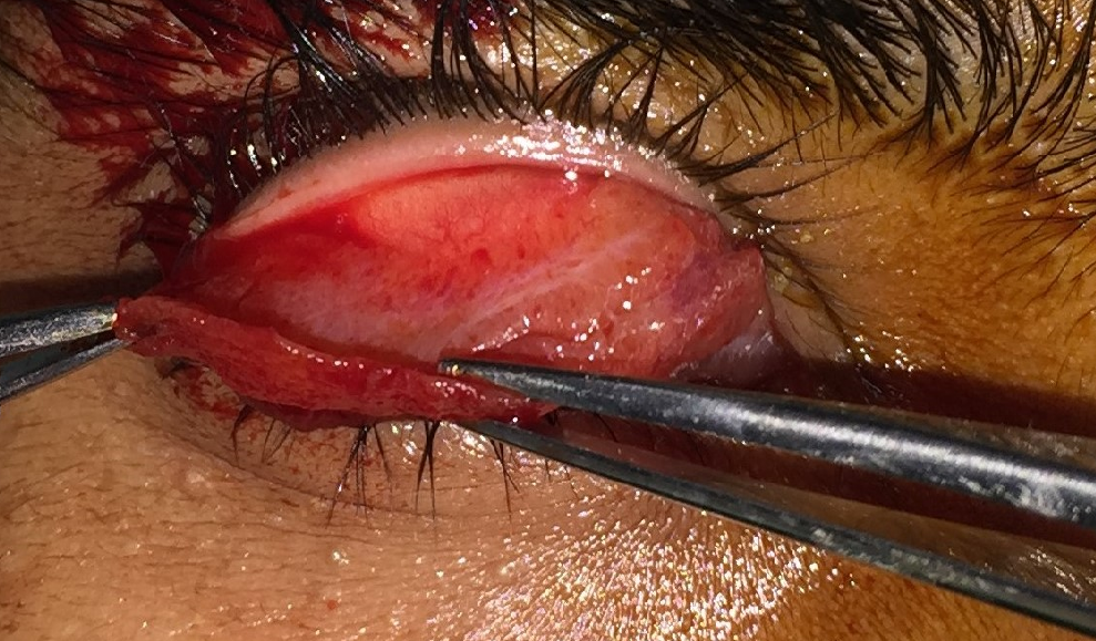
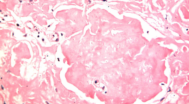
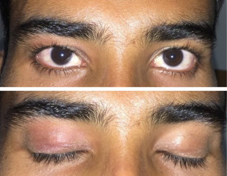
An 18 year old male presented to our institute with the complaint of drooping of right upper eyelid since four months and an unremarkable systemic and ocular examination except a moderate degree ptosis with levator action of 10mm. [Figure 1a,b] Posterior approach Levator Palpebrae Superioris (LPS) plication was planned. Per operatively after eversion of upper right eyelid the tarsus appeared hypertrophied and brittle. [Figure 2]. The abnormal looking tarsal tissue was excised and sent for histopathology (HP) and ptosis was corrected by levator plication. The histopathology (HP) of the excised tissue revealed eosinophilic acellular hyalinised material which had a positive apple green birefringence in polarising microscope with congo red staining, consistent with amyloidosis.[Figure 3] Investigation for systemic amyloidosis were negative. At one year follow up the lid height was satisfactory and patient had no ocular and systemic complaints. [Figure 4].
Discussion
Localized amyloidosis involving the tarsal conjunctiva and tarsus is a rare cause of chronic eyelid thickening and ptosis.1 There are various predisposing conditions and systemic involvement can affect various organs. Hence, localized disease warrants a thorough clinical evaluation and laboratory investigation.2 Absence of any skin changes on the eyelid or symptoms of ocular irritation, in our case, led us to an unsuspecting diagnosis of aponeurotic ptosis. Published literature mention recurrent haemorrhage, ocular irritation, multiple site involvement of conjunctiva, lid thickening and Levator muscle involvement as associated findings with ptosis.3 Our case had no signs and symptoms other than Ptosis at presentation highlighting the vast spectrum of presentation of amyloidosis .


