- Visibility 46 Views
- Downloads 13 Downloads
- DOI 10.18231/j.ijooo.2020.030
-
CrossMark
- Citation
Spontaneous conjunctival sudoriferous cyst- is it uncommon or underdiagnosed?
- Author Details:
-
Jasmitha B Rajashekar Reddy *
-
Darshan Bhatt
-
B Chandrasekaran
Introduction
Sudoriferous cysts, also known as hidrocystomas are rare cystic lesions arising from sweat glands distributed in different parts of the body, including the glands of Moll in the eyelids.[1] Congenital and post-traumatic orbital sudoriferous cysts are well documented in the literature. [1], [2] However orbital sudoriferous cysts of spontaneous origin in has been rarely reported.[3] Here we describe this unusual presentation.
Case Report
A 64 year old male patient presented to us with complaints of painless, gradually progressive diminution of vision in both eyes. There was no history of previous ocular surgeries or eye trauma. Patient underwent complete ocular examination, had BCVA of 20/80, N12 in the right eye and 20/60, N8 in the left eye. Slit lamp examination showed immature cataract in both eyes. On further detailed examination fullness of the lateral canthal angle was noted and on lateral displacement of the lateral canthal angle a single, well-defined, cystic swelling with smooth surface, blue to red in colour, measuring 8mm×8mm was noted arising from lateral forniceal conjunctiva, not involving the lateral canthal margin ([Figure 1]A). Ocular movements were full and painless. Rest of the ocular examination was normal. An X-ray skull and orbit imaging was done to rule out bony remodelling and orbital extension of the lesion, which showed normal study ([Figure 1]B). A diagnosis of simple conjunctival cyst was made and planned for cyst excision, followed by small incision cataract surgery(SICS) with PCIOL implantation on a later date. Patient underwent cyst excision under local anaesthesia, an in-toto excision of cyst was performed and sent for histopathological examination.
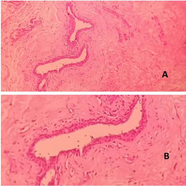
Gross examination showed a single grey-brown soft tissue measuring 8mm X 6mm X 2mm. Histopathological examination showed a cystic space lined by cuboidal epithelium, surrounded by fibro-collagenous tissue and congested vessels ([Figure 2]A). Luminal cells showed abundant eosinophilic cytoplasm along with apical snouting and decapitation secretions into the lumen ([Figure 2]B). The diagnosis of sudoriferous cyst was made. Postoperatively no complications were observed, 1 week post cyst excision patient underwent SICS with PCIOL implantation.


Discussion
Sudoriferous cysts are rare, benign cystic lesions involving the sweat glands, most commonly seen in head and neck region.[4] There are two types of sudoriferous cysts, eccrine and apocrine. Eccrine hidrocystomas are small thin walled cysts which can occur as single or multiple lesions, predominantly seen in adult females, mostly seen in the peri-orbital region. Apocrine hidrocystomas are relatively larger in size and usually solitary, mostly seen in head and neck region, along the eyelid margin and canthi. [4], [5] These cyst tends to remain asymptomatic for a longer period of time until they attain certain size. Literature review showed that all such cysts reported are of large size ranging from 1.5 to 4cm, either post-traumatic origin or are of congenital origin with bony remodelling. [1], [2], [6]
Orbital sudoriferous cysts are rare and only few cases are reported in children. [2] Conjunctival sudoriferous cysts are much more rare with trauma being the most common cause. [1] Till date only 3 cases of spontaneous origin sudoriferous cysts are reported in adults. [7], [8]
Common differential diagnosis of conjunctival and orbital cystic lesions are simple conjunctival cyst, dermoid cyst, lacrimal duct cyst, implantation cyst, haemangiomas. [9] Our case is an adult male who presented with spontaneous origin, asymptomatic, solitary, cystic lesion, with no radiological evidence of bony remodelling. Histopathological features showed no dermal appendages or lacrimal ductal tissue, which ruled out above mentioned common differential diagnosis. Thus our case was diagnosed as spontaneous origin conjunctival sudoriferous cyst.
On a routine day to day practice, small conjunctival cysts are not scrutinised and evaluated histopathologically. Thus there is a possibility of underdiagnosis of such cysts. Although rare, sudoriferous cysts should be considered as one of the differential diagnosis of conjunctival cystic lesions and histopathology plays a vital role in the definitive diagnosis.
Source of Funding
None.
Conflict of Interest
None.
References
- Anjali Mehta, Aparna Rao, Apoorva Khanna. Sudoriferous cyst of the orbit of adult origin after trauma. India J ophthalmol 2008. [Google Scholar]
- J. F. Saunders. Congenital Sudoriferous Cyst of the Orbit. Arch Ophthalmol 1973. [Google Scholar]
- B Kim, N Y Kang. Apocrine hidrocystoma of the conjunctiva. Int J Ophthalmol 2012. [Google Scholar]
- Abdullah Alfadley, Khalid Al Aboud, Asma Tulba, Mohamed Mazen Mourad. Multiple eccrine hidrocystomas of the face. Int J Dermatol 2001. [Google Scholar]
- K Sarabi, A Khachemoune. Hidrocystomas-a brief review. Med Gen Med 2006. [Google Scholar]
- Bahram Eshraghi, Mohammad-Ali Abtahi, Seyed Ali Sonbolastan, Zahra Kasaie, Seyed-Hossein Abtahi. Presentation of massive orbital hidrocystoma at birth: case report and review of the literature. Eye Vision 2017. [Google Scholar]
- K Chin, P T Finger, C Iacob. High-frequency ultrasound imaging of peribulbar hidrocystomas. Optom 2003. [Google Scholar]
- B Kim, N Y Kang. Apocrine hidrocystoma of the conjunctiva. Int J Ophthalmol 2012. [Google Scholar]
- J A Shields, C L Shields. Atlas of orbital tumors. 1999. [Google Scholar]
