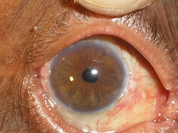
Introduction
Scleral fixation of IOL is the procedure of implanting a posterior chamber IOL in the sulcus in absence of posterior capsular support as in prior cataract extraction or Aphakia, Traumatic cataract, subluxation/dislocation of natural lens.
Various options for Secondary IOLs available are AC IOLs, Iris claw IOLs, Sutured IOLs, Glued IOLs and Sutureless Intrascleral IOLs. All of them have their own advantages and drawbacks.1
History
Scleral fixation of posterior chamber IOLs was first described by Malbran et al, in 19862
Stark et al,1989 placed IOL in ciliary sulcus by suturing haptic to sclera & iris. Lyle & Jin, 1992, did 11 & 5 o’clock fixation under triangular scleral flaps. Guinness et al, 1995 did 10.30 & 4.30 o’clock fixation with one straight & 1 curved needle. Uthoff et al 1998, 3 & 9 o’clock fixation was done.
Aims and Objectives
To study the outcome of 68 eyes who underwent sutured scleral fixation of posterior chamber IOL. Results in terms of Intraoperative difficulties, Postoperative complications and Visual recovery were noted.
Materials and Methods
68 eyes of 68 patients were subjected to sutured scleral fixation of posterior chamber IOL from Jun 2007 to Feb 2019 in Dept. of Ophthalmology of a teaching Government Institute.
The Ethics committee approved the protocol.
Indications of Scleral fixation of pcIOL- Inclusion criteria were
Prior aphakia of one eye with good Vision correction
PC rent or aborted primary IOL implantation
Subluxation of Lens
Traumatic Cataract with PC rent
Exclusion criteria were
Aphakia with BCVA of less than 6/60
Patients with aphakic glaucoma
With corneal pathology,
With inflamed eye, iritis, uveitis
With disorganized anterior segment/ organised scarring
With any fundus abnormality, retinal detachment / CME
68 Eyes of 68 patients were chosen for surgery by a single surgeon
Preoperative evaluation was meticulously done.
Best Corrected Visual Acuity, Routine examination of eye and adnexa, Slit lamp examination in detail to look at Limbus for incision, Corneal edema, scarring, Anterior chamber inflammation, Pupil and Iris- reaction to light, peaked, irregular pupil, or rubeoisis iridis, Status of posterior capsule remnants, Cortex, Vitreous Face and Fibrosis if any.
Fundus examination for CME or RD. followed by Keratometry and A Scan, and IOL power calculation. Intraocular Pressure measurement and Sac Syringing.
Timing of Scleral Fixation Surgery was decided as
Primary or Secondary- minimum 4 wks. after first surgery to take care of inflammation, hypotony and CME.
Procedure
After written consent and Peribulbar block, eye painted and draped, Speculum and Bridle suture applied, Conjunctival Peritomy performed, vessels cauterized, Triangular scleral flaps made 180 deg apart, 2 point (10-4, or 2-8 clock) 3 mm behind the limbus Pars Plana fixation, using One Straight and one curved needle10-0 Prolene suture. The straight needle was fed into a bent 26G needle and brought out. The suture loop brought out through the scleral tunnel incision with Mc pherson’s forcep, cut and then tied to the routine one piece pcIOL haptics at the highest point with 3 throws of tight knots. Posterior chamber IOL was then introduced behind the Iris taking care to avoid suture entangling. Scleral flaps covered the suture knots were sutured with 10-0 nylon.
Lewis technique was employed.
Selection of IOL and suture
Specifications of Suture supported IOLs 6-6.5 mm optic, 13-14 mm length
We used routinely available IOLs without eyelets, because these were available at our Government setup.
Suture used was10-0 prolene with one straight and one curved needle. The same suture needle was used to suture under the scleral flaps. Scleral flaps and scleral tunnel incision was sutured with 10-0 nylon.
Postop. Steroid and antibiotics e.d. given for 4 wks. Follow Up Was done on Day 1, wk 1,2,4, 3mths and 6 mths, and yearly thereafter.
At each visit Vision, Slit Lamp examination and Fundus was examined.
We had a long term followup of about 5-10 years.
Results
Timing of surgery
Primary surgery was done in 9 cases. Whereas we preferred Secondary surgery after 4 weeks in 59 (86.76%) cases.
Introperative difficulties
Hypotony made operating difficult in Primary surgery in 4 cases. Bleeding from Iris, ciliary body was seen in 2 cases. Suture entangling was seen in 2 cases. Suture breakage on one end was encountered on the table and the procedure needed to be repeated.
Postoperative complications
Hyphaema was seen in 2 cases. Immediate postop. Iritis was noted in 4 cases and was treated. CME was seen in 2 patients. RD was seen in 1 patient of traumatic Cataract after 1 month of surgery. Suture erosion, Decentration of IOL or Endophthalmitis was not seen.
Discussion
Options for Secondary IOL
A/c IOL
Iris claw IOL
Scleral fixated IOL- Sutured
Glued IOLs
Sutureless Intrascleral IOLs
Each has its own Advantages and Disadvantages.
Secondary A/C IOLs 3, 4
Advantages being- Easy to implant, Easy to explant and require less surgical time.
Complications being- Corneal Endothelial decompensation, edema., UGH syndrome, CME and poor Visual Acuity.
Complications
Central circular pupil required, Pseudophakodonesis, Decentration/ dislodge, Uveitis, Corneal Decompensation. Procedure is technically difficult.
Advantages being
IOL is placed closer to nodal point, Better optical properties, Less Psudophakodonesis, Less risk of Pupillary block Glaucoma, Less risk of Corneal Decompensation or UGH syndrome. Could be technically difficult and Time consuming.
Complications would be Hyphaema, RD.
Uhtoff Teichmann in Germany study in 624 patients, Observed that BCVA improved in 92% cases, Complications were IOL decentration in 1.9%, Suture erosion 17.9%, CME 5.8%, RD 1.4%,Vitreous Hmz 1%.5
In a Korean study by Shin W.J.in 7 eyes reported it to be safe method. (1 case had macular degeneration)6
Stem MS The riskes associated with sutured SFIOL surgery include the potential for postop lens dislocation, lens tilt, suprachoroidal or vitreous hmz, RD and endophthalmitis. The rates may vary based on the surgeon, patient circumstances and the technique used to affix the IOL to the svclera. In general, the complications arise from suboptimal suture placement. 7
In a 2015 study by Cavallini of 13 eyes with long term follow up from 5-10 yrs. found only 2 eyes with minimal lens decentration and this didn’t affect vision.8
Ma DJ studied ciliary sulcus and pars plana fixation and concluded using the pars plana for the iol haptics on pciol transscleral fixation is as effective as and safer than using ciliary sulcus. 9
Mahmood SA studied 70 eyes, and reported average BCVA improved to 6/12 with complications like transient IOP elevation(36%), IOL tilt(7.1%),CME (5.7%) Vit. Hmg(2.9%), hyphema(2.9%)uveitis(1.4%), and RD (1.4%).similar to our study.10
Luk ASW assessed the long term outcome and complication profile of SFIOL in a cohort of 104 eyes of chinese pts.72% improved final postoperative visual acuity.24% had postop. Complications. suture breakage with lenssubluxation occurred in 1.9% eyes.11
Mc Allistot AS, Hirst LW, 82 eyes studied with 72% improved corrected distant Visual acuity.12
Agarwal A described fibrin glue assisted sutureless pciol implantation with good results in 10 eyes.13
Hoffman, Scharioth, Yamane have recently advised various techniques like scleral pockets or scharioth scleral tunnels or Flanges for securing the sutures or IOL haptics., but these seem to have a learning curve and require special instruments, sutures and IOLs.14, 15, 16
Our study results were comparable to these other case series.
Visual outcome of these pts. with scleral fixated IOLs has been excellent. 92.5% patients had BCVA 6/18 - 6/6.
Precise location of anchoring points of lens at pars plana was important for preventing complications like bleeding from ciliary body, iris .
Scleral flaps to prevent Suture erosion & Endophthalmitis.
Proper technique and Tightening suture to prevent lens decentration and tilt.
Meticulous Anterior Vitrectomy to prevent Uveitis & CME.
No special IOL, or special instrument or expenses required.


