Introduction
The prevalence of pterygium in India ranges from 9.5% to 13%. 1 More than 50% of pterygium recurrences have been found to occur by 4 months after excision and nearly all (97%) by the first year. 2 Among the surgical treatments for pterygium, conjunctival limbal autograft is the surgery of choice with reduced recurrence rate. 3 Autograft is held in situ with the help of sutures or glue. Fibrin glue helps in decreasing post operative pain, surgical time and recurrence rate which is 5.3% 3 versus 13.5% 4 with suture and these heal with minimal inflammation. The major concern include the cost and since fibrin adhesives are prepared from pooled donor sources, there is a small but finite risk of infections such as hepatitis and HIV but there are no such reported infections.
Patient’s own blood is used as a bioadhesive in pterygium surgery and the recurrence rate was found to be similar to fibrin glue. 5 It is more cost effective with no risk of transmission of infection. The drawback of this technique is complications like graft displacement, loss of graft and graft retraction. The present study was conducted to know the complications with conjunctival limbal autograft surgery using patient’s own blood as adhesive for graft.
Materials and Methods
This is a prospective, cross sectional, time bound study conducted at a tertiary teaching institute in North Karnataka between June 2018 to Dec 2019. A total of 52 patients were included in the study between 25 years to 65 years with primary nasal pterygium and head of pterygium > 1mm into the cornea.
Exclusion criteria included (a) patients with history of previous ocular surgery or trauma or allergy (b) history of systemic diseases like diabetes mellitus or bleeding disorder (c) history of glaucoma, moderate to severe dry eyes, dacryocystitis, HIV or HBsAg.
Approval from Institutional Ethical Committee and written informed consent from each patient was taken. All the data was compiled and analysed using SPSS Software Version 19.
Preoperative Ophthalmic Evaluation included uncorrected and best corrected visual acuity, slit lamp examination, tonometry and fundoscopy. Photographic documentation was done before and after pterygium surgery. Grading of pterygium was done using Chao et al6 staging system for pterygium according to the extent of the pterygium.
Grade 1: pterygium just crossing the limbus.
Grade 2: pterygium crossing the midway between limbus and pupillary margin.
Grade 3: pterygium crossing the pupillary margin.
Routine preoperative investigations like blood sugar, urine routine, haemogra, and ECG were done.
All the surgeries were done under peribulbar anaesthesia following aseptic techniques and sterile draping. Superior rectus bridle suture was not placed. After placing the wire speculum, the neck of the pterygium dissected, lifted and avulsed from the corneal surface. Gentle scraping done to smoothen the corneal surface. Subconjunctival tissue under the body of the pterygium was excised including superior and inferior margins and the resultant bare sclera measured using Castroviejo calipers. 1mm oversize superior limbal conjunctival epithelium was retrieved avoiding buttonholing or tenon’s fascia. The limbal edge of the graft was carefully positioned at the host limbal tissue edge. Small haemorrhages seen under the graft were tamponaded with direct compression using nontooth forceps until hemostasis was achieved. The stabilization of the graft was tested centrally and on each free edge to ensure firm adherence to the sclera for 5 minutes before giving antibiotic steroid injection in inferior fornix and eye patched for 24 hours. All the surgeries were done by 2 surgeons who had good experience of pterygium excision with conjunctival autograft with suture technique.
Post operatively after 24 hours, the eye was assessed under slitlamp for graft adherence, symptoms or any other complications and were advised topical moxifloxacin, loteprednol and lubricating eye drops. Patients were followed up on 7th day, 1st month and 4th month. Any recurrence was noted i.e.,reappearance of fibrovascular growth at the site of previous excision extending beyond the limbus onto the cornea. Any intraoperative or post operative complications were noted.
Results
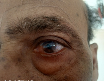
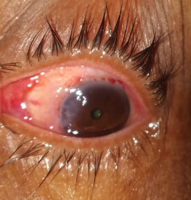
A total of 52 patients with primary pterygium were operated using sutureless, glueless conjunctival limbal autograft after pterygium excision. There were 30 males and 22 females with highest incidence among 35 to 55 years age group (78.85%). Majority of patients had grade 2 (76.9%) pterygium and most of them were farmers by occupation while most common indications for surgery were visual problems(48.07%) and cosmetic(40.38%) reasons.
Out of 52 eyes, graft remained in place in 49 patients. In 2 patients there was mild recession of the graft on canthal side while in 1 patient there was recession from the superior margin. None of the patient had displaced graft. 1 patient had pyogenic granuloma while in 12 patients there was nebular corneal opacity in the region of head of pterygium.
Table 1
Profile of Pterygium operated patients
| S. No. | Patient Profile | Total No |
| 1 | Total | 52 |
| 2 | Male | 30 |
| 3 | Female | 22 |
| 4 | Rural | 42 |
| 5 | Urban | 10 |
| 6 | Grade of pterygium type 1 | 8 |
| 7 | Grade of pterygium type 2 | 40 |
| 8 | Grade of pterygium type 3 | 4 |
Table 2
Total Numberof Patients with Symptoms / Complications on 1st, 7th and 30th Post Operative days
Discussion
Pterygium excision with conjunctival limbal autograft is well proven surgical procedure for pterygium with respect to minimal recurrence rate. The issues basically revolve around use of graft site, graft size and graft adherence. We used superior bulbar conjunctival epithelium as graft tissue after ruling out probability of glaucoma in selected patients with primary pterygium. Graft size of 0.5 mm larger size compared to bare sclera area was taken and placed with limbal to limbal orientation. The graft was gently tucked in (underneath the upper and lower conjunctival borders of excised pterygium) and gently pressed. The graft can be held in place by sutures or fibrin glue which have inherent side effects. Using patients own blood for adherence of graft has minimized disadvantages like post operative discomfort, cost, availability and time.
Cut and paste method technique for pterygium surgery using fibrin glue advocated by Koranyi et al3 reported that this technique was time saving and easy to learn. In our series of cases too we did cut and paste technique and used patients own blood at operating site as adhesive instead. Our technique avoids use of foreign materials such as suture and glue associated with increased inflammation, infection and hypersensitivity reactions. This technique is also cost effective. De wit et al7 in 2010 presented a case series describing simple technique of using a sutureless and gluefree method to fix conjunctivolimbal autograft using patients own blood containing fibrin at the bare sclera site. After placing the graft over pterygium excised site, we waited for 5 minutes for graft adherence as compared to study by Das gupta et al8 who waited for 10 minutes. It is now well accepted that, if once the graft stays in place for the first 24 to 48 hours, it will stick back. 3 patients in our study had graft dehiscense. This may be due to ocular movements or occasional rubbing by patients or inclusion of tenon’s in the graft.
1 patient had pyogenic granuloma during 7th day post operative visit which was treated with topical prednisolone drops. After 1 month we performed resurgery to excise the granuloma. This may be attributed to more tissue handling while excising the subconjunctival tissue of pterygium and also while placing the graft. 12 patients had mild corneal haze (nebular) over area covered by head of pterygium. This may be due to long lasting history of pterygium deeply embedded into corneal stroma.
Graft edema was a common complication 45 patients (86.53%) in our study while in a study by Shreesha kumar9 graft edema was observed in 52.04%. Graft edema completely subsided at the 7th day follow up. Subconjunctival hemorrhage and congestion was seen in 20 patients and it resolved completely by 1st month follow up. This might be due to absence of use of cautery and purposeful retention of blood under the graft.
None of the patient had recurrence at 4th month followup. Our study included only 52 patients as it was a timebound study. Short term followup and small sample size are the limitations in our study. Any surgeon well versed with conjunctival autograft technique using sutures can easily convert to this technique with almost no learning curve. Whether this technique should be considered as an ideal procedure for pterygium needs to be verified by multicentric large trials and however one would find difficult to perform this technique using topical anaesthesia.
Conclusion
With the current technique there are no suture related complications or glue related disadvantages. Complications will be less if one can perform meticulous dissection of tenon’s tissue from the conjunctival graft and recipient bed, minimal tissue manipulation, accurate orientation of the graft and holding onto the graft placement site for atleast 5 minutes for adherence.


