- Visibility 387 Views
- Downloads 82 Downloads
- Permissions
- DOI 10.18231/j.ijooo.2021.021
-
CrossMark

Introduction
Cryptophthalmos, meaning “hidden eye” is a rare ophthalmological presentation, first described by Zehender and Manz in 1872. It is an autosomal recessive inherited disorder and may occur as an isolated ophthalmological presentation or in association with various systemic disorders as in Fraser Syndrome or MOTA syndrome. Cryptophthalmic eyes, representing a fundamental failure in ocular development, have a poor prognosis for visual function, even if the retina is present. Cryptophthalmos has been classified into three morphological variants: complete, incomplete and abortive. Complete cryptophthalmos refers to a condition where the anterior portion of the eye is completely enveloped by a sheet of skin, that extends from the forehead to the cheek. The eyebrow, eyelid and associated adnexal structures are also absent, with the underlying eye being microphthalmic or staphylomatous, sometimes even normal. Partial cryptophthalmos, on the other hand, has eyelids covering the temporal portion of the eye normally, while the nasal part of the eyelid fold is absent, thereby leaving a sheet of skin lining the nasal side of the ocular surface encroaching the cornea. The anterior segment structures in such cases are normally formed, with shallow fornices, and such eyes have fair visual prognosis. Abortive cryptophthalmos, also known as congenital symblepharon, is an entity where the upper eyelid is partially or completely absent the lower eyelid being normal. [1], [2], [3], [4], [5]
Case Presentation
A 12 day neonate was brought to us at the Regional Institute of Ophthalmology, a unit of Motilal Nehru Medical College, Prayagraj, with complete cryptophthalmos in right eye and abortive cryptophthalmos in the left eye. He is the third living child, born to a 28yr old mother. The child was delivered at 9 months period of gestation through caesarean section in a local hospital. There is no history of delayed cry, any ICU admissions or any other pre- or peri-natal complications. The birth weight of the child was 2.30 kg with no post- natal complications.
The child is born to a couple in a non-consanguinous marriage. They have 3 living children together, out of which 2 suffer from cryptophthalmos. Two of their children have died shortly after birth due to post-natal complications, the details of which are not known. Both of them suffered from bilateral cryptophthalmos, as described by the parents. There is no history of any maternal genitourinary infections or systemic diseases during pregnancy. There is also no history regarding exposure to radiation or drug use during this or former pregnancies. The mother is however, anaemic. There is no history of similar, or any other familial, or genetic disorders in the immediate or distant family.
The child has a normal head circumference. On examination, the left eye was completely covered by skin. The eyebrows, eyelids, eyelashes were completely absent with no palpebral aperture. Spontaneous globe movements can be detected under the skin, with a soft eyeball on palpation.
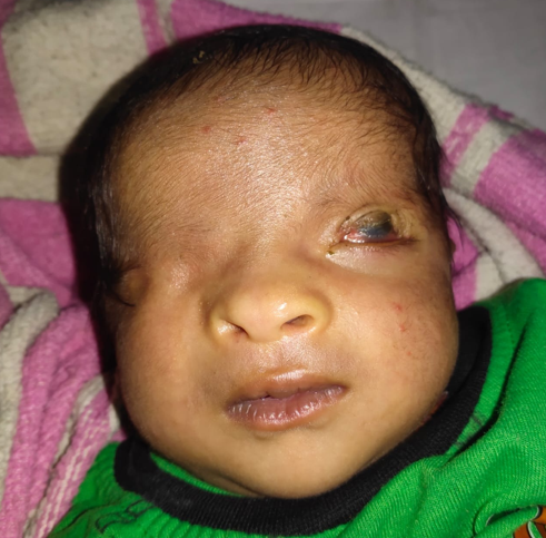
The left eye has a palpebral aperture, with well-formed lower eyelid with eyelashes, with upper eyelid coloboma. There are shallow fornices and conjunctivalisation and keratinisation of the cornea superiorly, probably a result of exposure keratopathy since birth. The left eyebrow is absent.
The child has a depressed nasal bridge. There is no umbilical herniation or genitourinary abnormalities. There are no other congenital defects of the fingers, ears or respiratory system.
B- scan ultrasonography of the orbits show the presence of a normally shaped right eye, 15 mm anterio-posterior axial length, with a shallow anterior segment and a probably cataractous lens with a normal posterior segment. The left eye has an axial length of 15.7 mm with normally formed anterior and posterior segments. No staphyloma is visible.
Discussion
Most cases of cryptophthalmos have been in association with Fraser syndrome. Isolated cryptophthalmos is rare, 27 cases seen so far.16 of these cases were sporadic and 11 familial. Familial cases occurred in three families and demonstrated vertical transmission. However, no such history of similar disease has been found in the parents’ families. Fraser Syndrome, first described by George Fraser in 1962, is a rare autosomal recessive disorder, characterised by cryptophthalmos, syndactyly, and abnormalities of the respiratory and urogenital tract. Pathogenesis of the disease is not yet known, though maternal deprivation of vitamin A and role of retinoids have been suggested in its etiology. The separation of eyelids occurs by a process of controlled necrosis of palpebral tissue between 17-18 weeks of gestation. A defect in the gene responsible for this programmed cell death may also be a factor. [6], [7], [8], [9]
In such cases, pre-natal screening and genetic counselling remains the only viable option. Bilateral orbital reconstruction with corneal grafting and anterior vitrectomy and lid reconstruction has been described in few cases but the visual deficit persists due to clouding of the corneal grafts, though the surgery may provide cosmetic benefits. We did not plan surgery in this patient because of the refusal of the parents to consent, given the poor visual prognosis post- surgery. Surgery can be attempted later in life to provide cosmetic improvement to the patient.
This patient has isolated, non familial, unilateral cryptophthalmos along with normal physical and mental milestones and normal hearing. No other associated malformations were identified.[10], [11]
The patient was prescribed eyedrop tobramycin, chloramphenicol+ polymyxin B eye ointment and lubricating eyedrops along with syrup Vitamin A to aid in corneal healing and the poor visual prognosis was explained to the parents.
Source of Funding
No financial support was received for the work within this manuscript.
Conflict of Interest
The authors declare they have no conflict of interest.
References
- Farooqui. Isolated BL Crytophthalmos. Nepal J Ophthalmol. 2016;8(16):186-204. [Google Scholar]
- Coulon P, Lan PT, Adenis JP, Verin P. Bilateral complete cryptophthalmos: Illustration with a case. Review of the literature. J Fr Ophthalmol. 1994;17:505-12. [Google Scholar]
- Kanhere S, Phadke V, Mathew A, Irani S. Cryptophthalmos. Indian J Pediatr. 1999;66(5):805-8. [Google Scholar] [Crossref]
- Thomas IT, Frias JL, Felix V, Leon LSd, Hernandez RA, Jones MC. Isolated and syndromic cryptophthalmos. Am J Med Genet. 1986;25(1):85-98. [Google Scholar] [Crossref]
- Kulkarni ML, Sureshkumar C, Venkataraman V. Syndromic and Isolated Cryptophthalmos. Indian Pediatr. 1995;32:1112-5. [Google Scholar]
- Kabra M, Gulati S, Ghosh M, Menon P. Fraser-Cryptophthalmos syndrome. Indian J Pediatr. 2000;67(10):775-8. [Google Scholar] [Crossref]
- Fraser GR. Our genetic load: A review of some aspects of genetic variation. Ann Hum Genet. 1962;25:387-415. [Google Scholar]
- Baijal GC, Agarwal ML, Chaurasia BD. Unilateral cryptophthalmos. Indian J Ophthalmol. 1981;29:41-2. [Google Scholar]
- Saal H, Traboulsi E, Gavaris P, Samango-Sprouse C, Parks M. Dominant syndrome with isolated cryptophthalmos and ocular anomalies. Am J Med Genet . 1992;43(5):785-8. [Google Scholar] [Crossref]
- Francois J. Malformative syndrome withcryptophthalmos. Acta Genet Med Gemellol. 1969;18:18-50. [Google Scholar]
- Harris MJ, McLeod MJ. Eyelid growth and fusion in fetal mice. Anat Embryol. 1982;164(2):207-20. [Google Scholar] [Crossref]