- Visibility 166 Views
- Downloads 31 Downloads
- Permissions
- DOI 10.18231/j.ijooo.2024.032
-
CrossMark

An analytical study of ophthalmological manifestations in hematological malignancies in a tertiary care centre, India
- Author Details:
-
 Raja Ayyakutty Muni *
-
Seema Gurusamy
Abstract
Aim: The objective of the study was to describe the ophthalmological manifestations associated with various haematological malignancies at a tertiary care centre.
Materials and Methods: This analytical cross-sectional study was conducted with 60 patients diagnosed with various types of haematological malignancies, including leukaemia, lymphomas, and multiple myeloma. Patients who had ocular media opacities which precluded fundus view like dense corneal opacity, cataracts, absolute glaucoma, and systemic conditions like hypertension, and diabetes were excluded from this study. All 60 patients underwent a comprehensive ocular examination and the collected data were analysed using IBM SPSS software version 25.
Results: Out of the 60 patients with haematological malignancies, 27 (45%) exhibited ocular features. Ocular involvement was more common in males (63%) and in the paediatric age group (1-10 years) (100%). Ocular findings were prevalent in leukaemia patients especially with chronic myeloid leukaemia showing the highest incidence (80%), compared to lymphomas and multiple myeloma. The most common ocular manifestations were in the posterior segment, such as retinal haemorrhages and Roth spots, observed across all age groups. Neurological involvement, specifically III and VI cranial nerve palsy, were also notable. At the time of presentation, the majority of patients with ocular features had good visual acuity, ranging from 6/6 to 6/9 (43%).
Conclusion: With advancements in diagnostic and therapeutic methods, the survival rates for patients with haematological malignancies have considerably improved. This has led to an increase in the variability of ocular presentations. Consequently, routine ocular examination is essential at the time of diagnosis of haematological malignancies to prevent vision-threatening complications and improve the overall quality of life for these patients.
Introduction
The hematological malignancies prevalent in India are leukemia, multiple myeloma, and lymphoma, with a higher incidence among males compared to females.[1] The common types of hematological malignancies are Acute Lymphocytic Leukemia (ALL), Chronic Lymphocytic Leukemia (CLL), Acute Myeloid Leukemia (AML), Chronic Myeloid Leukemia (CML), Hodgkins Disease (HD), Non-Hodgkins Lymphoma (NHL), and Multiple Myeloma (MM). [1], [2]
Ophthalmic manifestations are seen in approximately 90% of the patients with hematological malignancies.[3], [4] This could be attributed to either direct infiltration by the leukemic cells into the eye or secondary to pancytopenia, anemia, intracranial hypertension, hyperviscosity, radiotherapy or chemotherapy.[5], [4] The ocular manifestations could involve either the anterior segment or the posterior segment. The common anterior segment features are ptosis, proptosis, dry eye, conjunctivitis, subconjunctival haemorrhage, scleritis, sclerokeratitis, corneal infiltrates, anterior uveitis, and pseudohypopyon.[6], [2] Posterior segment manifestations include posterior uveitis, vitreous haemorrhage, Roth spots, pre-retinal haemorrhage, and papilledema. Third and sixth cranial nerve palsies are the common neuro-ophthalmic manifestations. [2], [4]
Ocular involvement is most commonly seen in chronic myeloid leukemia in approximately 68% - 76% of cases.[3], [4] Liebreich first described Leukemic retinopathy in 1860.[7], [8] The ocular involvement in leukemia was reported as 90% by Osama Badeeb. In lymphoma, the ocular involvement is very uncommon and can present as retinal vasculitis, proptosis, and uveitis.[9], [10] In multiple myeloma, there is direct infiltration of the ocular tissues with neoplastic cells which causes proptosis, corneal deposits, and ciliary body cysts. [11], [12]
Materials and Methods
This analytical cross-sectional study was conducted at Government Medical College, Palakkad, Kerala, from November 2018 to October 2022.
Inclusion criteria
Adults aged ≥ 18 years diagnosed with leukemia, lymphoma, or multiple myeloma.
Pediatric patients diagnosed with hematological malignancies.
Exclusion criteria
Individuals with the following conditions were excluded
Dense corneal opacity
Cataract
Diabetes
Systemic hypertension
A total of 60 patients who met the inclusion criteria and were referred from oncology department to the ophthalmology outpatient department with hematological malignancies for ophthalmology evaluation were included in this study. Demographic details, including age, gender, occupation, and comprehensive medical history, such as type, duration and treatment details of malignancy were noted. After obtaining informed consent from all the adult patients and parents of pediatric patients as per the Declaration of Helsinki, all patients underwent complete ocular examinations like visual acuity assessment using Snellen’s chart, intraocular pressure measurement with Goldman’s applanation tonometer, anterior segment examination with Topcon slit lamp biomicroscopy, fundus examination using +90D lens as well as direct and indirect ophthalmoscopy. Fundus photography was done for selected cases. Blood smear investigations were done on all patients to determine the type of hematological malignancy and bone marrow examination was also done for selected patients. All data were entered into a Microsoft Excel sheet and analyzed using IBM SPSS software version 26.
Results
Out of the sixty patients with hematological malignancy, 27 (45%) were found to exhibit ocular features. In this study, the age of the patients with hematological malignancy varied from 1 year to 79 years, and the ocular features were also presented in a wide range of age groups. ([Table 1]) Among those patients with ocular features, posterior segment findings were more common than the anterior segment involvement among all age groups. Out of the sixty patients included in this study, 38 were males and 22 were females. Ocular findings were present in 19 of the 38 males and 8 of the 22 females. Among the ocular features, anterior segment findings were present in 4 males and 2 females. Neurological involvement was found in 3 males and 1 female. Posterior segment findings were present in 12 males and 5 females, indicating that posterior segment involvement was more prevalent than anterior segment or neurological involvement.
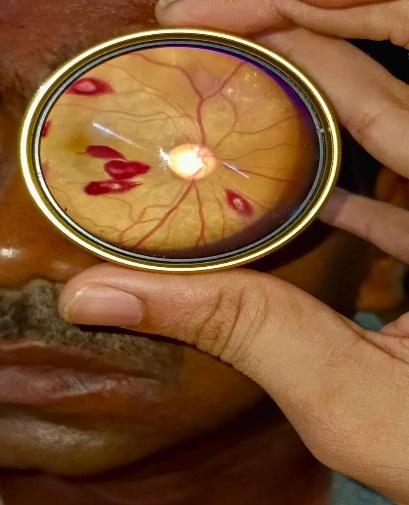
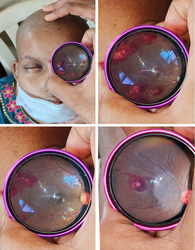
In this study, ocular manifestations were more common in males (63%) compared to females (37%). The gender groups, posterior segment features were predominant. This difference follows the higher incidence of hematological malignancies in males.[11] Chronic myeloid leukemia was present in 5 patients, out of which 4 had ocular features (80%) ([Figure 1]). Chronic lymphoid leukemia was present in 5 patients, with only 1 patient had ocular signs (20%). Among 11 patients with Acute lymphoblastic leukemia, 6 had ocular signs (55%) (Figure 2), and among 7 patients with acute myeloid leukemia, 5 patients had ocular manifestations (71%). Among 11 patients with Hodgkin’s disease (HD), 3 patients had ocular manifestations (27%). Among 13 patients with multiple myeloma (MM), 6 patients had ocular features (46%), and non-Hodgkin’s lymphoma (NHL) was present in 8 patients, out of which 2 patients (25%) had ocular features. Among the ocular manifestations the posterior segment findings were higher than the anterior segment findings in leukemia patients except for AML, which had an equal incidence and CML, which had one case of anterior segment findings, one case of neurophthalmic manifestation and two had posterior segment findings. Among the various ocular manifestations, posterior segment findings were most common, like retinal haemorrhages and Roth spots in 5 patients each followed by pre-retinal haemorrhages in 2 cases in our study. Next in frequency was nerve palsies, including 3rd and 6th nerve palsies with ptosis and diplopia in 4 cases. Next in order were vitreous haemorrhage and posterior uveitis, with two cases each. Other ocular manifestations such as proptosis in 2 cases, anterior uveitis in 2 cases, papilledema, sclerokeratitis, and dry eyes, were observed in one case each. ([Table 2])
Among 60 patients with 120 eyes, about 50 eyes (42%) of 25 patients had a fairly good visual acuity ranging from 6/6 – 6/9. 26 eyes (22%) of 13 patients had a visual acuity between 6/12 – 6/18, and 24 eyes (20%) of 12 patients had a visual acuity ranging from 6/24 – 6/36, respectively. The remaining 20 eyes (16%) of 10 patients had an acuity ranging from 6/60 to PL+. One patient with Hodgkin lymphoma presented with right eye axial proptosis with cavernous sinus infiltration and total retinal detachment. He had a vision of only perception of light. There was a 2 ½ years old male child with right eye axial proptosis and poor visual acuity, limited to perception of light. Another 2-year-old male child had right eye vitreous hemorrhage with visual acuity of counting fingers close to the face.
|
Age (years) |
Number of patients |
Ocular features |
Anterior Segment including neurological manifestations |
Posterior Segment manifestations |
|
1-10 |
7 |
7 |
2 |
5 |
|
11-20 |
9 |
3 |
1 |
2 |
|
21-30 |
10 |
5 |
3 |
2 |
|
31-40 |
12 |
3 |
1 |
2 |
|
41-50 |
12 |
3 |
1 |
2 |
|
51-60 |
5 |
2 |
1 |
1 |
|
61-70 |
3 |
2 |
0 |
2 |
|
>70 |
2 |
2 |
1 |
1 |
|
Total |
60 |
27 |
10 |
17 |
|
Type of hematological malignancy |
Total no. of cases (n=60) |
Ocular features |
Ocular manifestations |
|
|
Value (n=27) |
Percentage |
|||
|
AML |
7 |
5 |
71% |
• Nerve palsy |
|
|
|
|
|
• Anterior uveitis |
|
|
|
|
|
• Sclerokeratitis |
|
|
|
|
|
• Roth spots |
|
|
|
|
|
• Papilledema |
|
CML |
5 |
4 |
80% |
• Proptosis |
|
|
|
|
|
• Nerve palsy |
|
|
|
|
|
• Retinal hemorrhage |
|
|
|
|
|
• Roth spot |
|
ALL |
11 |
6 |
55% |
• Anterior uveitis |
|
|
|
|
|
• Posterior uveitis |
|
|
|
|
|
• Vitreous Haemorrhage |
|
|
|
|
|
• Pre retinal haemorrhage |
|
|
|
|
|
• Retinal haemorrhage |
|
|
|
|
|
• Roth spots |
|
CLL |
5 |
1 |
20% |
• Retinal haemorrhage |
|
MM |
13 |
6 |
46% |
• Proptosis |
|
|
|
|
|
• Nerve palsy |
|
|
|
|
|
• Dry eye |
|
|
|
|
|
• Posterior uveitis |
|
|
|
|
|
• Vitreous hemorrhage |
|
|
|
|
|
• Retinal haemorrhage |
|
HD |
11 |
3 |
27% |
• Nerve palsy |
|
|
|
|
|
• Roth spots |
|
|
|
|
|
• Retinal haemorrhage |
|
NHD |
8 |
2 |
25% |
• Roth spots |
|
|
|
|
|
• Pre retinal haemorrhage |
Discussion
Prevalence of ocular involvement in hematological malignancies, according to various studies varies from 24% to 70%.[13], [14], [15] In our study, out of 60 patients, 27 (45%) patients had ocular involvement. Osama Badeeb et al. reported that ocular involvement in leukemias is 90%.[16] Elise Torczynski et al. revealed primary ophthalmic leukemia infiltrate in 3% of their patients., secondary ophthalmic findings in 39%, and ocular changes related to leukemia treatment in 20%.[16], [13]
In our study, ocular features were observed in the age group between 1-79 years. Among the 60 patients, 16 patients were under 20 years old, 34 were between 20–50 years old, and 10 were aged between 51–80 years old, which was similar to the Osama Badeeb et al. study.[16] The posterior segment findings were predominant in all the age groups, particularly among those aged between 1-10 years.
The time interval between leukemia diagnosis and the onset of ocular manifestation was ≤ 6 months in 11 patients and 24 to 60 months in six patients. The lowest interval reported was 1 month, observed in 3 patients.[14], [15]
In our study, ocular manifestations were common in adults (74%) than children (26%). A similar study conducted by Reddy et al. revealed that the eye changes were common in adults (49.1%) compared to children (16.5%).[17]
In this study, posterior segment findings were the most common manifestations which includes retinal and pre-retinal hemorrhages, vitreous hemorrhages, Roth spots, sub-retinal hemorrhages, papilledema, retinal detachment, and posterior uveitis, constituting about 63% of the ocular manifestations. The anterior segment findings were dry eye, sclerokeratitis, and anterior uveitis, constituting 22% of the ocular manifestations, and neurological manifestations including 3rd and 6th nerve palsies accounted for 4 cases (15%). This contrasts with a study conducted by Moll et al. reported that anterior segment findings were more common than posterior segment findings.[16], [17]
In this study, Cranial nerve palsy secondary to CNS infiltration was observed in 3 patients. Two patients with ALL had proptosis. In this present study, one case of papilledema, no case of optic nerve infiltration were noted. In mulitiple myeloma, cysts of the ciliary body and retinal vascular lesions were the most common ocular manifestations. Matano et al. reported a patient with AML who developed leukemic hypopyon.[18] In this study, one patient with acute myeloid leukemia had bilaterally established papilledema.[18] Saenzfrances et al. reported that a case of bilateral papilledema secondary to chronic lymphocytic leukemia.[19], [20] In our study, one case of anterior uveitis and one case of unilateral rhegmatogenous retinal detachment that occurred in patient with acute lymphocytic leukemia, which was similar to Atchaneeyasakul et al. reported a case of anterior uveitis in a patient with relapsing ALL. Among the 60 patients in our study group, 8 had non-Hodgkins lymphoma, with 2 had ocular features (25%), which was similar to Kazuhiro Yamada et al.'s reported a case of primary B-cell non-Hodgkins lymphoma with anterior uveitis.[21] Hodgkins disease was present in 11 patients, with only 3 patients had ocular manifestations (27%), which was similar to the Valenzuela J et al. study.[22]
13 patients with multiple myeloma 6 had ocular features (46%), which was similar to the Shoji MK et al. study.[23] Francois Thoumazet et al. reported two cases of multiple myeloma presenting with orbit and muscle involvement and distal bony erosion.[24] Pradhan S. et al. reported a case of multiple myeloma with bilateral tearing due to infiltrative lesions of the lacrimal sac.[25]
In this study, among 60 patients with 120 eyes, nearly 42% (n=50 eyes) of patients presented with hematological malignancies had visual acuity ranging from 6/6–6/9. 22% (n=26 eyes) had 6/12–6/18 vision. About 20% (n=24 eyes) had 6/24–6/36. 16% (n=20 eyes) of the patients had a vision of 6/60 or worse. In this study, poor vision is due to pre-retinal hemorrhage at the macula, vitritis, axial proptosis, vitreous hemorrhage, cavernous sinus infiltration, and sixth nerve palsy, which was similar to the study conducted by Hendrik AM et al.[26]
Conclusion
Our study concluded that ocular involvement is very high in leukemias, especially chronic myeloid leukemia cases and among pediatric patients. Posterior segment involvement, such as pre-retinal hemorrhage and roth spots were commonly associated with various types of ocular malignancies. As ocular involvement may arise before the systemic diagnosis, during the course or relapse period, we recommend that ocular examinations be routinely conducted at the time of diagnosis of all cases of hematological malignancies.
Our study recommends the importance of interdisciplinary collaboration between hemato-oncologists and ophthalmologists for comprehensive ocular screening to prevent vision-threatening complications and improving overall patient quality of life.
Source of Funding
None.
Conflict of Interest
None.
References
- . WHO. WHO Classification of Tumours of Haematopoietic and Lymphoid Tissues. . 2009. [Google Scholar]
- Thareja J, Minj A, Samal P, Panigrahi P. Ophthalmic manifestations in hematological malignancies: An observational study from a tertiary care health center in Eastern India. Indian J Ophthalmol. 2024;72:659-63. [Google Scholar] [Crossref]
- Rossi C, Buizza A, Alessio G, Borselli M, Taloni A, Carnevali A. Ophthalmic Manifestations in Patients with Blood Malignancies. Hematol. Rep. 2024;16(2):193-203. [Google Scholar]
- Bouazza M, Youssefi H, Bouanani N. Ocular manifestations in hematological disorders. Cureus. 2022;14(8). [Google Scholar] [Crossref]
- Zachariah G, Idiculla T. A prospective study of ophthalmic manifestations of common haematological disorders. Int J Acad Med Pharm. 2024;6(2):521-4. [Google Scholar]
- Suresh K, Sampath R, T. Ocular manifestations in hematological disorders. SRMC. 2011;4:1-4. [Google Scholar]
- Aisenberg A. Historical review of lymphomas. Br J Haematol. 2000;109(3):466-76. [Google Scholar]
- Ilo O, Adenekan A, Alabi A, Onakoya A, Aribaba O, Kehinde M. Ocular manifestations of leukaemia: A teaching hospital experience. Niger Postgrad Med J. 2019;26(4):205-10. [Google Scholar]
- Skarsgård L, Andersson M, Persson M, Larsen A, Coupland S, Stenman G, et al. Clinical and genomic features of adult and paediatric acute leukaemias with ophthalmic manifestations. BMJ Open Ophthalmol. 2019;4(1). [Google Scholar] [Crossref]
- Soman S, Kasturi N, Srinivasan R, Vinod K. Ocular manifestations in leukemias and their correlation with hematologic parameters at a tertiary care setting in South India. Ophthalmol Retina. 2018;2(1):17-23. [Google Scholar]
- Menon L, John E, Deepthi R, Shaji A. Prevalence of ocular manifestation in haematological malignancies-A clinical study. IOSR J Dent Med Sci. 2018;17(6):22-8. [Google Scholar]
- Talcott K, Garg R, Garg S. Ophthalmic manifestations of leukemia. Curr Opin Ophthalmol. 2016;27(6):545-51. [Google Scholar]
- Orhan B, Malbora B, Bayar S, Avcı Z, Alioğlu B, Özbek N. Ophthalmologic findings in children with leukemia: a single-center study. Turk J Ophthalmol. 2016;46(2):62-7. [Google Scholar]
- Koshy J, John M, Thomas S, Kaur G, Batra N, Xavier W. Ophthalmic manifestations of acute and chronic leukemias presenting to a tertiary care center in India. Indian J Ophthalmol. 2015;63(8):659-64. [Google Scholar]
- Pandharinath J, Chandrakant S, Pandharinath J. Ophthalmic manifestations of common haematological disorders. J Evol Med Dent Sci. 2014;3(42):10510-6. [Google Scholar]
- Badeeb O, Tashkandi I, Omar A, Anwer M, Farwan K, Dabbagh A. Ocular Leukemia in King Abdulaziz University Hospital - Jeddah. Ann Saudi Med. 1995;15(3):222-6. [Google Scholar]
- Reddy S, Jackson N, Menon B. Ocular involvement in Leukemia - A study of 288 cases. Ophthalmologica. 2003;217(6):441-5. [Google Scholar]
- Salloukh NE, Hage D, Bashshur A, Kheir W. Early Ophthalmological Manifestations of Acute Myeloid Leukemia: Current Perspectives. Clin Ophthalmol. 2022;16:2119-27. [Google Scholar] [Crossref]
- . Saenz Frances F et al; ocular manifestations in chronic lymphocytic leukemia. Arch SOC ESP ophthalmol. 2007;82:303-6. [Google Scholar]
- Buchan J, Mckibbin M, Burton T. The prevalence of ocular disease in chronic lymphocytic leukaemia. Eye. 2003;17:27-30. [Google Scholar] [Crossref]
- Valenzuela J, Echegaray J, Dodds E, Kurup S, CLowder, Ondrejka S. Ophthalmic Manifestations of Hodgkin Lymphoma: A Review. Ocul Oncol Pathol. 2021;7(6):381-9. [Google Scholar]
- Shoji M, Chen Y, Topilow N, Khzam RA, Dubovy S, Johnson T. Orbital Involvement in Multiple Myeloma. Ophthalmic Plast Reconstr Surg. 2023;39(4):347-56. [Google Scholar]
- Pennisi M, Berchicci L, Miserocchi E, Mussetti A, Cacioppo V, David A. Ocular disorders in multiple myeloma patients: cross-sectional study of prevalence and association with treatment. Leuk Lymphoma. 2019;60(2):477-82. [Google Scholar]
- Chin K, Kempin S, Milman T, Finger P. Ocular manifestations of multiple myeloma: Three cases and a review of the literature. Optometry. 2011;82(4):224-30. [Google Scholar]
- . Hendrik et al; ocular manifestations of hematological malignancies. Am J Hematol. 1979;7(4):389-94. [Google Scholar]